Fourth International Workshop @ MICCAI
Cancer Prevention, Detection, and IntervenTion
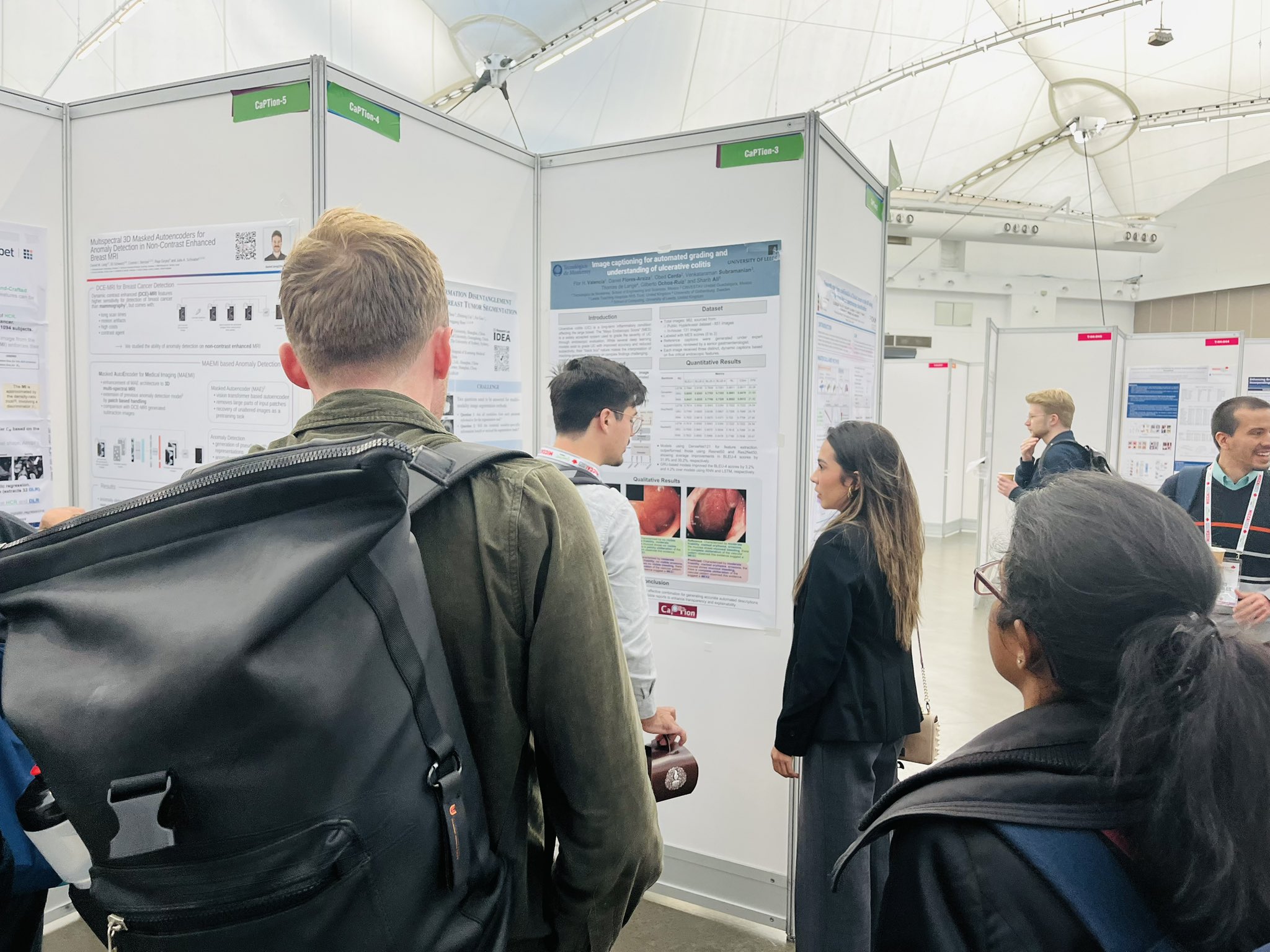
CaPTion
Submit Your Paper

Fourth International Workshop @ MICCAI
While computational methods in medical imaging have enabled us to detect and assess cancerous tumors and assist in their treatment, early detection of cancer precursors provides us with an opportunity for its early treatment and prevention. The survival rate of cancer is still low, and largely depends on the affected organ and how early it is diagnosed.
The variable nature of the disease in different patients and the diverse imaging acquisition types involved for quantification of disease and treatment demands robust method designs. It is therefore critical to develop generalizable methods as part of a holistic early cancer detection ecosystem.
The workshop will invite researchers in the field of medical imaging around the central theme of data-driven cancer detection and treatment, and strives to address the challenges that are required to be overcome to translate computational methods to clinical practice through well designed, generalizable (robust), interpretable and clinically transferable methods.
New this year: We will be including an exciting panel discussion on "Tackling medical imaging challenges in early detection, prevention and treatment of cancer."
Learning algorithms for lesion detection in medical images, staging, risk assessment, prediction of cancer outcome.
Image fusion, multi-modal registration, detection, segmentation, and tracking, computer-guided interventions, augmented reality.
Big imaging data analysis, active, semi & self-supervised learning, meta-learning, federated learning, LLMs, and continual learning.
New predictive visual biomarker discovery in medical images, tumor data signatures, personalized cancer treatments, genomics and radiomics.
Identifying new evaluation metrics or gold standards, sample size standardization, biases, uncertainty estimation, and image simulation techniques.
All papers should be formatted according to the Springer Lecture Notes in Computer Science (LNCS) templates.
We recommend submission up to 8-pages and 2-pages of references.
We adhere to a double-blind peer review process. Please follow the MICCAI 2026 anonymity guidelines when preparing your initial submission.
Submission Portal: OpenReview Website
Accepted papers will be published in a joint proceeding with the MICCAI 2026 conference via Springer LNCS.

TU/e, Eindhoven

University of Leeds, UK

AASTMT, Egypt

University of Oxford, UK

National Univ. of Singapore

University of Leeds, UK

University of Leeds, UK
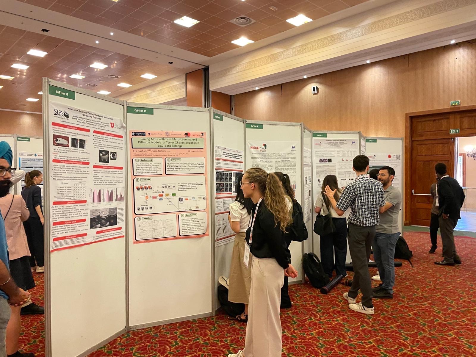
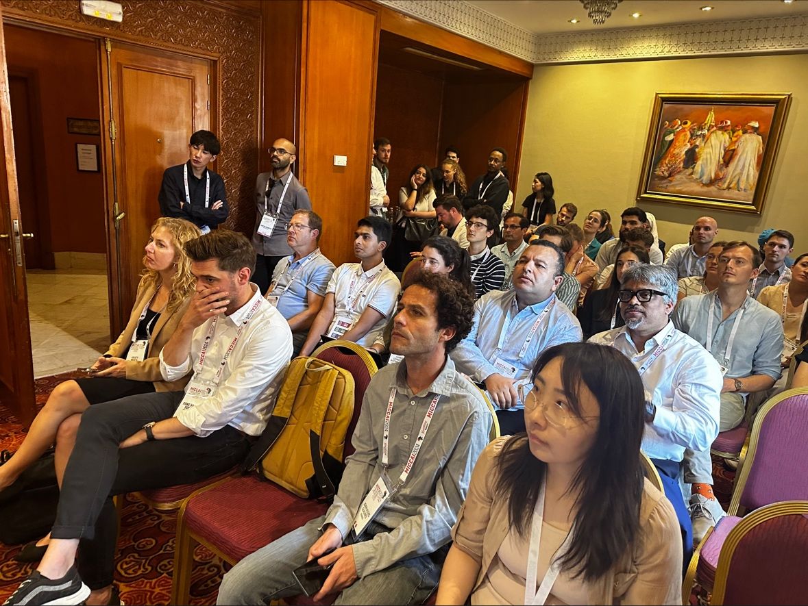
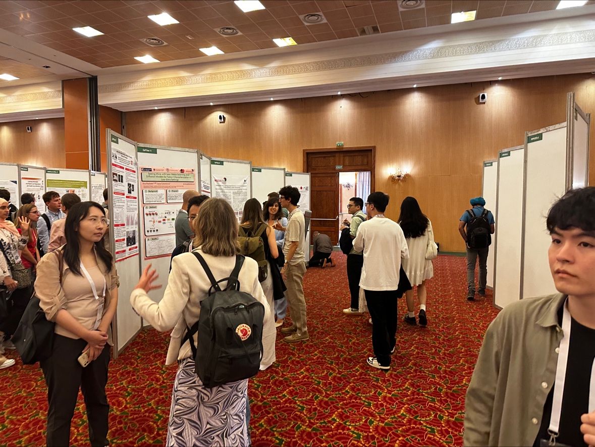
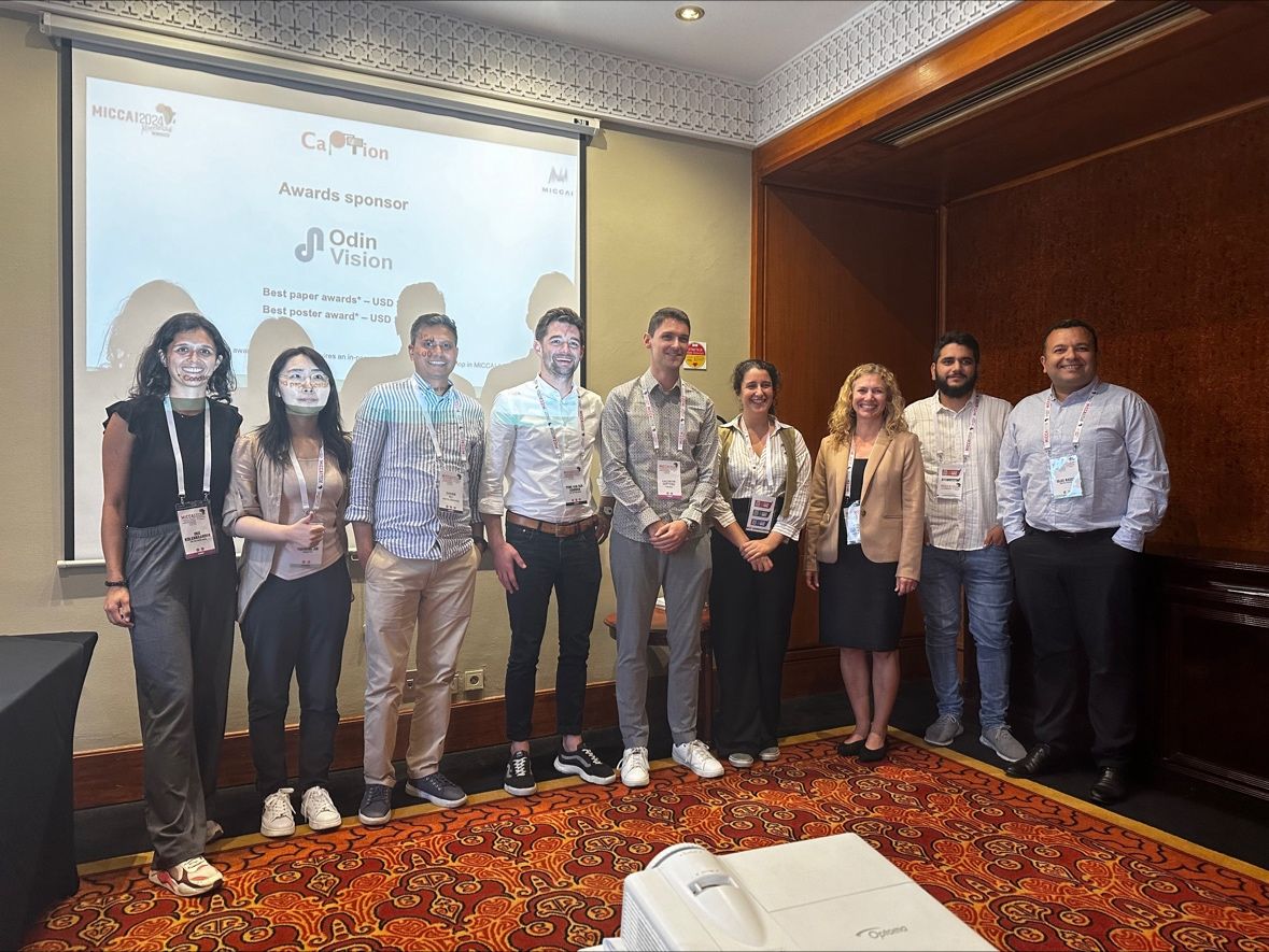
A look back at previous successful CaPTion workshops, keynotes, and poster sessions.