News!!!
Workshop will be conducted on 12th (13:30 Vancouver, Canada time) : Workshop Schedule CaPTion@MICCAI2023
Submission open till **14th July 2023**
Submissions opens on 2nd May 2023 : CMT for CaPTion@MICCAI2023
Submit your 8-10 page MICCAI format paper here (please read submission instructions)
Workshop Description
While computational methods in medical imaging have enabled us to detect and assess cancerous tumors and assist in their treatment, early detection of cancer precursors provides us with an opportunity for its early treatment and prevention. The survival rate of cancer is still low, and largely depends on the affected organ and how early it is diagnosed. The variable nature of the disease in different patients and the diverse imaging acquisition types involved for quantification of disease and treatment demands robust method designs. It is therefore critical to develop generalizable methods as part of a holistic early cancer detection ecosystem -- including different data analysis methods for various modalities and optimizing cross-modality fusion for improved detection and prognosis.
The workshop invites researchers to submit their work in the field of medical imaging around the central theme of early cancer detection, and it strives to address the challenges that are required to be overcomed to translate computational methods to clinical practice through well designed, generalizable (robust), interpretable and clinically transferable methods. Most current methods are developed on retrospective data that do not guarantee good representation of daily clinical procedures (e.g. domain gap). Through this workshop, we aim to identify the new ecosystem that will enable comprehensive method validation and reliability of methods, setting up a new gold standard for sample size and elaborate evaluation strategies to identify failure modes of methods when applied to real-world clinical environments.
Workshop themes
Early detection and diagnosis of cancer: learning algorithms for lesion detection in medical images, staging, risk assessment, prediction of cancer outcome (in terms of life expectancy, survivability, progression, treatment sensitivity)
Image-guided therapy for cancer treatment: Image fusion, multi-modal registration, detection, segmentation, and tracking, computer-guided interventions, augmented reality for tumor delineation
Real-world data exploration for cancer prediction: Big (imaging) data and analytics, active learning, semi and self-supervised learning, model-agnostic meta-learning, federated learning for (sparsely labeled) multi-center data.
Cancer biomarkers: new predictive (visual) biomarker discovery in medical images, tumor data signatures, personalized cancer treatments, genomics and radiomics.
Clinically accepted evaluation methods: Identifying new evaluation metrics or gold standards (e.g., compared to widely used Dice or IoU metrics), sample size standardization, best practices for validation, image simulation and synthesis techniques to train more robust deep learning models for cancer detection.
Keynote speakers
 Dr Anne Martel is a Professor in Medical Biophysics at the University of Toronto, a Senior Scientist and Tory Family Chair in Oncology at Sunnybrook Research Institute, and a Faculty Affiliate at the Vector Institute, Toronto. Her research program is focused on medical image and digital pathology analysis, particularly on applications of machine learning for segmentation, diagnosis, and prediction/prognosis. In 2006 she co-founded Pathcore, a software company developing complete workflow solutions for digital pathology.
Dr Martel is an active member of the medical image analysis community and is a fellow of the MICCAI Society. She has served as board member of MICCAI and is currently on the editorial board of the journal Medical Image Analysis..
 Sir Michael Brady is an Emeritus Professor in the Departments of Oncology and Engineering Science at the University of Oxford, having retired as a Professorship in Information Engineering (1985-2010). He is also a Distinguished Honorary Professor of the Mohamed bin Zayed University of Artificial Intelligence in Abu Dhabi. He was awarded the IEE Faraday Medal for 2000, the IEEE Third Millennium Medal for the UK, the Henry Dale Prize by the Royal Institution in 2005, and the Whittle Medal by the Royal Academy of Engineering 2010. Mike was knighted in the New Year’s Honours list for 2003. Mike has founded companies, including ScreenPoint Medical (breast cancer), Volpara Health Technologies (breast cancer), Optellum (lung cancer), Perspectum (liver, pancreas cancer). He has served on the Early Detection and Diagnosis, and the Multidisciplinary, Committees of Cancer Research UK.
 Dr. Sravanthi Parasa is a practicing gastroenterologist and clinical researcher based at the Swedish Medical Center in Seattle. Dr. Parasa's research focuses on the intersection of epidemiology, biostatistics, and machine learning with a passion for augmenting patient care through the meaningful applications of Artificial Intelligence and digital technologies. She is an active member of GI society committees and serves on IEEE/engineering and computer science conferences and program committees, where she is a frequent invited speaker on the translation of digital and AI solutions to patient care. By partnering with world-renowned institutes, she has published several papers and guidelines in areas of high-fidelity risk prediction models, application of computer vision and natural language processing in the medical space.
Key technical themes
Medical image analysis methods targeted to the detection, diagnosis, treatment and/or monitoring of cancer that include a wide range of machine learning algorithms, workflows designed to improve patient outcome, data analysis and insights, and new imaging datasets.
Some key technical themes are listed below (not limited to):Learning algorithms & workflows (e.g., semi and self-supervised, active learning, metric learning, model-agnostic meta-learning, unsupervised learning, outlier detection)
Data and label efficiency (e.g., limited data problem, data imbalance problem)
Model robustness and generalisability (e.g., edge-AI, cloud-AI, quantized models for clinical application)
Explainability, fairness and data privacy (e.g., model calibration, saliency mapping and federated learning for multi-center data)
Important Dates
Paper submission begins: 2nd May 2023
Submission deadline: 30th June 14th July 2023 (final extension)
Paper decision notification: 12th 28th July 2023
Camera ready submission: 20th 7th August 2023
Workshop day: 12th October 2023
Submission
Accepted papers will be published in a joint proceeding with the MICCAI 2023 conference.
All papers should be formatted according to the Lecture Notes in Computer Science templates.
We recommend submission up to 8-pages and 2-pages of references (same as MICCAI main conference) for a double-blind peer review process
In addition, since the joint workshop has adhered to the double-blinded peer review process, we ask that you please follow the MICCAI2023 anonymity guidelines when preparing your intial submission.
Proceeding
Accepted papers will be published in LNCS as a separate CaPTion 2023 (MICCAI Workshop) , Singapore, 22 October 2023 proceeding
Call for Papers
Imaging themes (not limited to):
Optical imaging: Endoscopy, OCT, Hyperspectral imaging, opto-acoustics
CT/PET fusion, MRI
New imaging biomarkers
Multimodal imaging
Ultrasound
Clinical applications (not limited to):
Early cancer detection and diagnosis
Prognosis and prediction
Tumor characterisation, cancer staging
Longitudinal patient studies
Surgical data science
Digital histopathology
Phenotypic tumor correlation
Workshop Programme
| -Start- | -End- | -----Topic---- | --------Speaker----------- |
|---|---|---|---|
| 13:30 | 13:35 | Welcome (Introduction and objective of CaPTion) | Organisers |
| 13:35 | 14:15 | Keynote lecture #1 | Prof. Sir Michael Brady, FRS, FREng, FMedSci (University of Oxford) |
| 14:15 | 14:45 | Oral session #1 | Speakers: Van Linh Le, University of Bordeaux and Pablo Cesar Quihui-Rubio, Tec de Monterrey |
| 14:45 | 15:25 | Keynote Lecture #2 | Prof. Anne L Martel (University of Toronto, Canada) |
| ----- | ----- | ----- | ----- |
| 15:25 | 16:00 | Coffee Break & Poster Session | 11 posters |
| ----- | ----- | ----- | ----- |
| 16:00 | 16:15 | Oral session 2 | Speaker: Rebeca Vétil, Telecom Paris |
| 16:15 | 16:55 | Closing lecture | Dr. Sravanthi Parasa (Gastroenterologist, Swedish Medical Centre in Seattle, USA) |
| 16:55 | 17:00 | Closing remarks and Awards | Special Guest/Organisers |
Accepted Papers
Classification
Paper 1: A Deep Attention-Multiple Instance Learning framework to Predict Survival of Soft-tissue Sarcoma from Whole Slide Images
Van-Linh Le et al.
Paper 2: Towards Real-time Confirmation of Breast Cancer in the OR using CNN-based Raman Spectroscopy Classification
David Grajale et al.
Paper 3: Fully Automated CAD System for Lung Cancer Detection and Classification Using 3D Residual U-Net with multi-Region Proposal Network (mRPN) in CT Images
Anum Masood et al.
Paper 4: Image Captioning for Automated Grading and Understanding of Ulcerative Colitis
Flor Helena Valencia et al.
Detection and Segmentation
Paper 5: Multispectral 3D Masked Autoencoders for Anomaly Detection in Non-Contrast Enhanced Breast MRI
Daniel M. Lang et al.
Paper 6: Non-redundant Combination of Hand-Crafted and Deep Learning Radiomics: Application to the Early Detection of Pancreatic Cancer
Rebeca Vétil et al.
Paper 7: Assessing the Performance of Deep Learning-Based Models for Prostate Cancer Segmentation Using Uncertainty Scores
Pablo Cesar Quihui-Rubio et al.
Paper 8: MoSID: Modality-Specific Information Disentanglement from Multi-parametric MRI for Breast Tumor Segmentation
Jiadong Zhang et al.
Cancer/Early cancer Surveillance
Paper 9: Colonoscopy Coverage Revisited: Identifying Scanning Gaps in Real-Time
George Leifman et al.
Paper 10: ColNav: Real-Time Colon Navigation for Colonoscopy
Netanel Frank et al.
Paper 11: Modeling Barrett’s Esophagus Progression Using Geometric Variational Autoencoders
Vivien van Veldhuizen et al.
Organising committee
 |
 |
 |
|
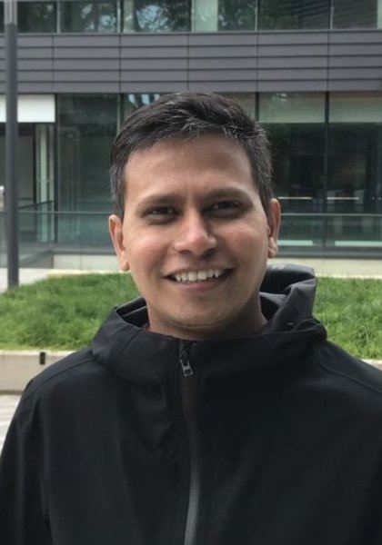
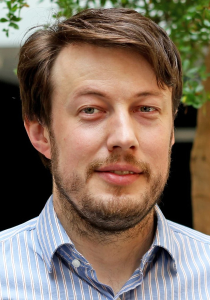
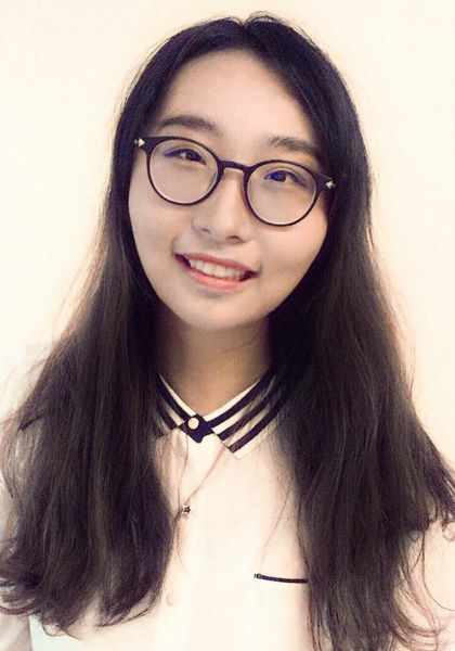
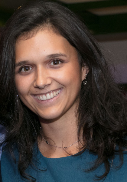
| Sharib Ali University of Leeds, UK |
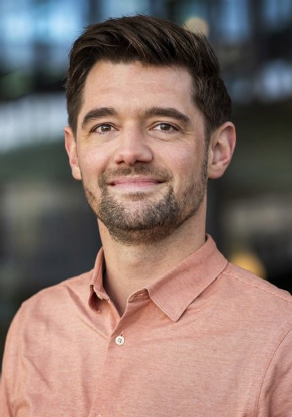
Fons van der Sommen TU/e, Eindhoven, The Netherlands |
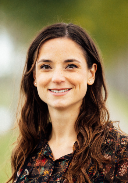
Maureen van Eijnatten TU/e, Eindhoven, The Netherlands |
|
 |
 |
 |
|
| Bartek Papiez University of Oxford, UK |
Yueming Jin National University of Singapore |
Iris Kolenbrander TU/e, Eindhoven, The Netherlands |
Student Representative
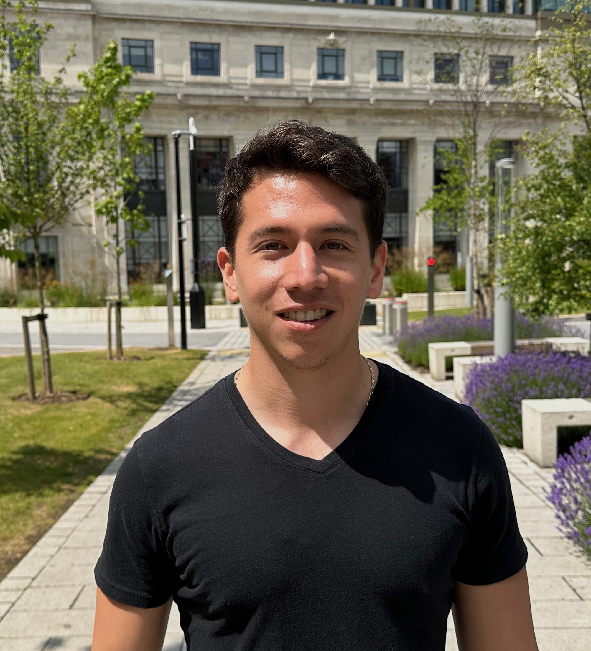 |
| Pedro Chavarrias Solano University of Leeds, UK |
Scientific Review Committee
| Christian Daul (Université de Lorraine, France) |
| Mariia Dmitrieva (Queen Square Analytics, UK) |
| Noha Ghatwary (University of Lincoln, UK) |
| Mark Janse (UMC Utrecht, The Netherlands) |
| Tim Jaspers (Eindhoven University of Technology, The Netherlands) |
| Carolus Kusters (Eindhoven University of Technology, The Netherlands) |
| Van Linh Le (University of Bordeaux, France) |
| Gilberto Ochoa-Ruiz (Tecnológico de Monterrey, Mexico) |
| Mansoor Ali Teevno (Tecnológico de Monterrey, Mexico) |
| Christiaan Viviers (Eindhoven University of Technology, The Netherlands) | Ziang Xu (University of Oxford, UK) | Jiadong Zhang (ShanghaiTech University, China) |
Sponsors
 "Satisfai" Satisfai Health Inc is a GI‐focused health technology developer that uses industry leading data and world class experts paired with artificial intelligence to provide real‐time decision-making support to physicians, with planned products covering the whole gastrointestinal tract. Our solutions deliver real-time analysis of medical imagery and provide clinicians with decision-making intelligence that dramatically improve patient outcomes.
Contact us
Please contact us for more details or if you are interested in sponsoring the event. Contact CaPTion team at: caption.miccai@gmail.com
Previous workshops
CaPTion Workshop @ MICCAI 2022 CaPTion Workshop @ MICCAI 2022
CaPTion2022 Springer Proceeding


